Extracellular vesicles
Observe EV heterogeneity, surface biomarkers, and corona dynamics with single-particle resolution
Capturing the true heterogeneity of extracellular vesicles
Extracellular vesicles (EVs) are premier candidates for non-invasive liquid biopsies and targeted drug-delivery vectors. However, characterising EVs and translating discovery into clinical realities is plagued by several major analytical bottlenecks. Many techniques require extensive isolation steps (such as ultracentrifugation or size-exclusion chromatography) to separate EVs from the background noise of plasma or lysates, potentially destroying fragile coronas that contain important biological information. These molecules are also inherently heterogeneous, from cargo variance to polydispersity, which can be challenging to quantify.
Solution-based single-molecule spectroscopy tracks individual particles as they diffuse through a focused laser volume, permitting differentiation between freely diffusing background proteins and intact vesicles based on size and brightness. This enables rigorous analysis of native EVs directly in unpurified serum, plasma, or cell lysates.
Fluorescence correlation spectroscopy (FCS) quantifies molecular size, polydispersity, and local viscosity at pM to nM concentrations. The addition of another colour permits cross-correlation (FCCS) that identifies the synchronised movement between two molecules, such as EV and cargo or two surface markers. Additionally, single-molecule Förster resonance energy transfer (smFRET) can probe the conformational states of surface receptors or study molecular crowding within the native lipid bilayer.
Unmask the true bionanoscale identity of extracellular vesicles
- Perform heterogeneity profiling by measuring the diffusion speed of vesicles directly in unpurified biofluids
- Track corona formation and dynamics in real time
- Quantify the co-diffusion of generic vesicle markers and disease biomarkers, identifying EVs that express both
- Distinguish EVs from free protein contaminants in cell lysates or serum
- Quantify surface biomarker density, counting the absolute number of specific receptors or cargo proteins present per vesicle
- Understand conformational changes of surface-bound proteins in response to environmental changes or ligand binding
- Assess EV integrity and cargo retention
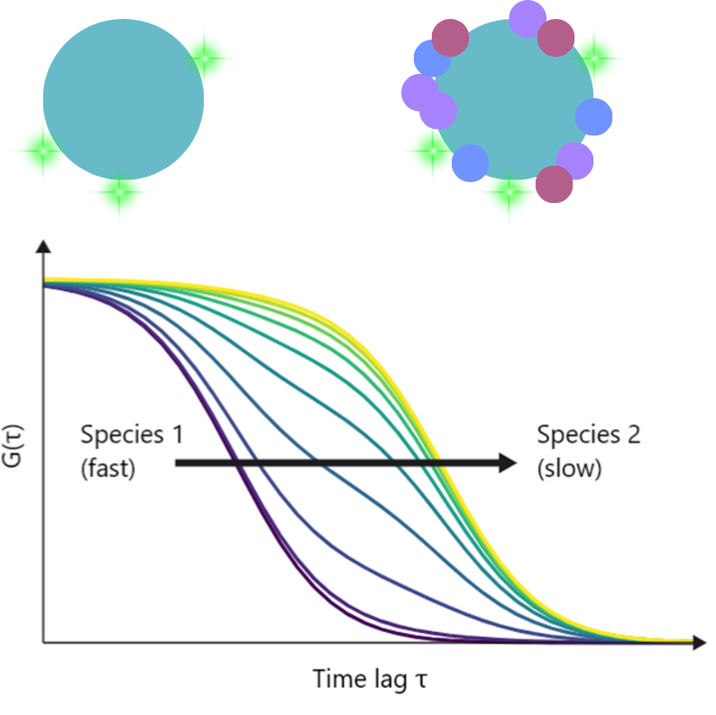
In the illustrative example shown below, EVs can be labelled with a dye and their diffusion analysed by FCS. When they are analysed in plasma, these vesicles begin to form coronas as proteins associate with their surfaces. This slows their diffusion, shifting FCS autocorrelation curves to the right. This figure is based on recent work by Musicò et al.
Extracellular vesicle resources


Extracellular vesicle FAQs
How can this technology analyse EVs in crude serum or lysates without purification?
Our confocal platform works by focusing a laser into a microscopic volume (less than one femtoliter) and tracking individual fluorescent particles. Because EVs can be labelled with fluorescent dyes, they can be differentiated from background proteins, even if they are also autofluorescent.
What are the benefits of studying unpurified EVs over purified samples?
Purification methods like ultracentrifugation or size-exclusion chromatography (SEC) apply mechanical stress that can lyse or deform fragile vesicles. More importantly, purification often strips away the biomolecular corona – the dynamic cloud of host proteins that associates with the vesicle in vivo.
How does FCS measure the biomolecular corona?
The biomolecular corona changes the physical size of the vesicle. As proteins associate with the vesicle surface, its hydrodynamic radius increases. By tracking the diffusion time of the vesicles via FCS, you can watch the autocorrelation curve shift, allowing you to quantify the formation and stability of the corona.
Can this technology identify which specific proteins make up the corona?
By labelling a suspected corona protein (e.g., an apolipoprotein) with one colour and the EV membrane with another, FCCS will measure the cross-correlation to inform what percentage of vesicles have recruited that specific protein into their corona.
Can we monitor the exchange or displacement of proteins within the corona?
Yes. Because our system monitors equilibrium dynamics in solution, you can perform displacement assays. For example, you can pre-form a corona with one labelled protein, introduce a competing unlabelled fluid or a different plasma environment, and use FCS/FCCS to watch the labelled proteins dissociate or be replaced in real-time.
How does the platform validate that a disease biomarker is present on EVs?
This is a major challenge for liquid biopsies, as free-floating tumour proteins can easily contaminate a sample. If a general EV marker (like CD63) is labelled with one fluorophore and the disease target with another, the EV can be analysed using FCCS. Biomarkers in solution will diffuse independently to the EV, and if the target is embedded in the vesicle, the two colours will produce a cross-correlation signal.
Can the system determine the absolute abundance of a biomarker per vesicle?
Photon Counting Histogram (PCH) analysis, alongside FCS, measures the molecular brightness of individual vesicles. Since the brightness is directly proportional to the number of fluorophores passing through the laser at once, you can calculate the absolute number of specific receptors or cargo proteins present on a single EV.
Can I differentiate between EVs and similar-sized lipoproteins (e.g., LDL/HDL)?
Yes. Physical sizing tools like NTA cannot distinguish an exosome from a lipoprotein of the same size. Our platform relies on molecular specificity via fluorescent labelling, meaning only molecules of interest will be analysed, irrespective of whether they are of a similar size to surrounding particles.
How can this technology assist in the development of engineered EV therapeutics?
For engineered EVs (such as those designed to deliver siRNA or small-molecule drugs), you need to know if your loading protocol was successful. By labelling the EV shell with one colour and the internal therapeutic cargo with another, FCCS can determine exactly what percentage of the vesicles are successfully loaded, and FCS can confirm if the engineering process altered the native size or stability of the vesicle.
Can I measure cargo leakage or shelf-life stability?
Absolutely. Because the system can distinguish between a slow-moving vesicle and a fast-moving, free molecule, a simple leakage assay is highly effective. If your therapeutic cargo leaks out of the vesicle over time or under certain conditions, you will see a distinct shift in the FCS curve from a slow, vesicle-bound diffusion profile to a fast, free-molecule profile, giving you a direct percentage of cargo retention.
"It has opened up new avenues of research because of the combined FCS, FCCS and single-molecule FRET capabilities. The high performance, stability and precision of the EI-FLEX instrument have really been a game-changer for experiments where sensitivity to picomolar concentration of biomolecules really matters."
Dr Steven Quinn, University of York

Other resources you might be interested in