The biological world exists across energy landscapes, involving transient intermediates, rate-limiting conformational shifts, and complex binding equilibria. In order to derive understanding from this energetic chaos, we have perfected observing biology in snapshots, from frozen or crystalline structures to averaged readouts of our cells fixed in a moment in time.
This has provided a wealth of information, but omits a key component of life, from the subcellular to the macroscopic: biology is dynamic.
To truly understand these systems, we need to view them as they are, in solution and in motion, observing individual molecules to find the rare intermediates that would otherwise be lost amongst the average.
In this article, we’ll explore three techniques (smFRET, FCS, and FCCS) that capture these vital in-solution dynamics, highlighting case studies where these methods revealed mechanistic insights, linking structure to function:
- How smFRET captures the global conformational changes induced by nucleotide binding of Rag GTPase and the allosteric impact of mutations in the nucleotide binding domain
- The use of smFRET and FCS to investigate the mechanism of action of two distinct antiviral drugs against SARS-CoV-2 RNA-dependent RNA polymerase
- How FCCS characterises ternary complex formation and kinetics, from cooperativity to stability
The solution-phase toolkit (smFRET, FCS, and FCCS)
Single-molecule FRET (smFRET), Fluorescence Correlation Spectroscopy (FCS), and Fluorescence Cross-Correlation Spectroscopy (FCCS) add the dynamic layer that brings static structures to life. By observing individual molecules or correlation events in solution, these tools reveal mechanisms of action (MOA), kinetic rates, and allosteric transitions that underpin biological processes.
smFRET: Mapping the conformational energy landscape
Single-Molecule Förster Resonance Energy Transfer (smFRET) is used to measure atomic-scale distances. By labelling two sites on a biomolecule with fluorescent dyes, the distance between them (and how this alters between conformational states) can be measured across a 3-10 nm range.
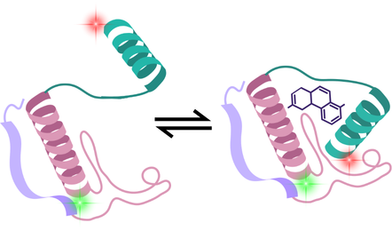
smFRET resolves distinct structural subpopulations within heterogeneous samples and captures conformational changes in response to ligand interactions or environmental changes, such as pH or salt concentration. With further analysis, the interconversion rates between conformations can also be calculated.
FCS: In-solution binding affinities and aggregation analysis
Fluorescence Correlation Spectroscopy (FCS) measures the diffusion of individual molecules through a femtolitre-scale volume. Given that smaller particles diffuse faster than larger ones or those bound to other biomolecules, size, binding affinity, and concentration can all be calculated from this data.
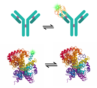
Therefore, FCS detects changes in molecular size caused by conformational expansions, oligomerisation, or ligand binding in real time. It also provides information on aggregation; due to the sensitivity of FCS, it can detect initial small oligomers before they form aggregates, providing insights into the early stages of aggregation.
FCCS: Dual-colour molecular co-trajectories
Fluorescence Cross-Correlation Spectroscopy (FCCS) takes FCS a step further. It uses two different-coloured lasers to determine whether two biomolecules are moving through the solution as a single unit. Cross-correlation of this signal will only occur when these two biomolecules are diffusing together.
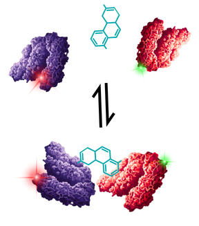
This provides a direct, solution-based proof of co-binding, making it ideal for understanding the stoichiometry of complexes and quantifying protein-protein interactions. Unlike methods where molecules must be tethered to a surface, FCCS measures binding entirely in solution, eliminating surface immobilisation artefacts, mass transport limitations, and steric hindrance. It excels at probing high-order, multi-component interactions, such as ternary complexes and molecular glues, under physiological conditions.
Case Study 1 – Uncovering how disease mutations alter allosteric mechanisms (smFRET)
Understanding how eukaryotic cells sense amino acids and trigger growth relies on decoding the rapamycin complex 1 (mTORC1) pathway and the action of Rag GTPase. When amino acids are abundant, the heterodimer adopts an active state that recruits mTORC1 to the lysosomal surface.
Rag GTPases are heterodimeric, undergoing global conformational shifts that reorient these heterodimeric subunits relative to one another.
Crystallographic and cryo-EM models have successfully captured snapshots of Rag complexes, but without seeing how these complexes move in solution, static structures alone cannot explain how point mutations outside of the active site drive disease.
Doxsey & Shen (2025) tackled this challenge by using single-molecule FRET (smFRET) to track the global conformational movements of individual Rag GTPase heterodimers in real time. By placing donor and acceptor fluorophores on different subunits, they observed the effect of nucleotide binding on the dynamic conformational changes of the protein1.

Figure 1 – smFRET detects dynamic conformational changes of Rag GTPase in response to nucleotide binding
Modified figure from publication by Doxsey and Shen, 2025 1 .
Of note, they saw that mutations in the nucleotide binding pocket influenced the global protein conformational state:
- Dual GDP binding of RagA-RagC(S75N) produced a pronounced mid-FRET population, which shifted further to a high-FRET state when GTP was added. This aligns with the observation that RagA-RagC(S75N) mutants drive forward mTORC1 signalling, as Rag GTPase is shifted towards the active conformation even when the incorrect combination of nucleotides is bound.
- The RagA(T21N)-RagC mutant remained in the open, inactive conformation regardless of the combination of nucleotides bound, in agreement with the observation that this mutation inhibits mTORC1.
To read more about this study and explore their smFRET experimental set-up in more detail, see our application note.

Figure 2 – Impact of nucleotide-binding domain mutations on global Rag GTPase conformations
FRET efficiency histograms for mutated Rag GTPase and overlays between mutant and wildtype for each condition: A) Unbound RagA-RagC(S75N), B) RagA-RagC(S75N) with dual-bound GDP, C) RagA-RagC(S75N) with GDP and GTP, D) Unbound RagA(T21N)-RagC), E) RagA(T21N)-RagC with dual-bound GDP, F) RagA(T21N)-RagC with GDP and GTP
Modified figure from publication by Doxsey and Shen, 20251 .
Case Study 2 – Tracking real-time catalysis and drug inhibition (smFRET and FCS)
Understanding how viral enzymes synthesise nucleic acids, and how nucleotide analogue drugs interrupt that process, is a cornerstone of antiviral drug discovery. To explore these dynamic questions, Groves et al. developed an in vitro solution-phase platform combining smFRET and FCS to investigate the mechanism of action of two SARS-CoV-2 RNA-dependent RNA polymerase (RdRp) inhibitors: a nucleotide competitive inhibitor (remdesivir) and a non-nucleotide inhibitor (sumarin)2.
Here, they formed a reporter RNA hairpin labelled with donor and acceptor fluorophores that would demonstrate a high-FRET state when unextended, and a low-FRET state when the hairpin had been extended by RdRp.
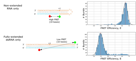
Figure 3 – smFRET reporter system using an RNA hairpin to demonstrate inhibition of viral RdRp by antivirals
Modified figure from publication by Groves et al., 2025 2
This model was well suited to capturing inhibition of RdRp by sumarin, which prevents RNA template binding and disrupts RNA entry into the active site. A clear dose-response could be seen relative to the extent of low- to high-FRET states, and therefore extended reporter RNA molecules.

Figure 4 – smFRET data showing sumarin inhibition of RdRp extension of labelled RNA
FRET efficiency histograms across increasing sumarin concentrations, ranging from 0 to 400 µM.
Modified figure from publication by Groves et al., 2025 2
Remdesivir does not show a clear dose-response, with little change in the ratio of FRET states as the concentration increases. FCS experiments show that RdRp stalls on RNA transcripts in low nucleotide conditions, and the authors proposed that the antiviral activity of remdesivir may be delayed in this context, whereby inhibition occurs as a result of inefficient incorporation of UTP opposite remdesivir molecules in the template strand, acting as a potential secondary mechanism of action.
You can explore this study further in our application note here.
Case Study 3 – Quantifying ternary complex cooperativity and kinetic stability (FCCS)
As the therapeutic landscape grows ever more complex, drug discovery pipelines can no longer rely on simple binding assays to determine whether a candidate is worth progressing. This is especially true for modalities such as ternary complexes, where strong binding candidates may not necessarily induce targeted protein degradation. It is crucial, therefore, to understand the link between initial binding, cooperativity and stability, and how these factors collectively influence the productivity of resultant ternary complexes.
Current assays for ternary complex characterisation often involve complex deconvolution of binary data, or surface-based experiments, which are known to introduce artefacts. In a recent publication from Exciting Instruments, we establish FCCS as a solution-phase approach for measuring the affinity, cooperativity, and kinetic stability of higher-order protein assemblies3.
These data focus on a clinically important model system: the simultaneous targeting of the extracellular domain of HER2 by two distinct monoclonal antibodies, trastuzumab and pertuzumab. While both therapeutics are known to bind distinct epitopes on HER2 and exhibit synergistic clinical efficacy in cancer therapy, the precise biophysical mechanisms driving their ternary assembly in solution had remained contested.
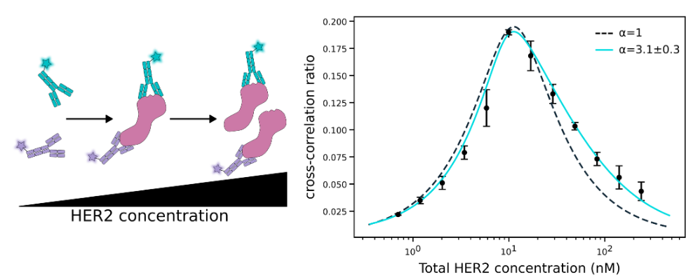
Figure 4 – FCCS data produces a characteristic hook plot for ternary complexes
As HER2 concentration increases, ternary complex formation also rises until binary species begin to dominate. FCCS data directly captures this hook effect, while also identifying a positive cooperativity value of 3.1.
Figure adapted from Mueller et al., 2026 3
Using FCCS in a microplate-compatible workflow, we recorded cross-correlation signals arising exclusively from co-diffusing, fluorescently labelled proteins, permitting ternary complex characterisation:
- Direct detection of ternary assembly: Because FCCS cross-correlation relies strictly on physical co-diffusion through a tiny confocal volume, the assay quantified true ternary complex formation in solution without surface immobilisation artefacts or rigid spatial requirements
- Quantifying positive cooperativity (α): By titrating HER2 against labelled trastuzumab and pertuzumab, the FCCS readouts revealed positive cooperativity (α). The binding of the first antibody to HER2 enhances the binding affinity of the second antibody, demonstrating an allosteric crosstalk between distinct epitopes
- Measuring enhanced kinetic stability (koff): Dissociation experiments carried out by FCCS proved that the fully assembled HER2–trastuzumab–pertuzumab ternary complex has greater stability (exhibiting a significantly slower off-rate) than either binary antibody–antigen interaction alone
For more information on this experiment, see the application note here.
Conclusion
Biological function is inherently dynamic, comprised of fluctuating energy landscapes, allosteric population shifts, step-by-step kinetic transitions, and multi-component assembly equilibria. In order to understand these processes, our experimental techniques should, therefore, also be dynamic, bringing static structures to life and pulling individual heterogeneity out of the noise of the average.
By probing macromolecules in their native-like environments, solution-phase single-molecule and correlation techniques provide the resolution required to complete the picture:
- smFRET reveals how allosteric networks and disease-causing mutations alter global dynamic equilibria, or can act as a reporter system for RNA extension
- FCS quantifies diffusion, hydrodynamic shifts, and binding affinities, all without ensemble averaging
- FCCS directly measures multi-body binding stoichiometry, positive cooperativity (α), and kinetic stability (koff) in solution, bypassing the steric and mass-transport limitations of surface-bound assays
If you would like to find out more about how you can apply in-solution dynamics to your own research, discover a wealth of information in our resource library, or get in touch with one of our application scientists today.