Protein aggregation
Identify early-stage aggregates in complex biological systems
Characterise protein aggregates – from biotherapeutic stability to neurodegenerative biomarkers
The ability to detect and characterise protein aggregation is crucial in both drug development and disease detection. In drug development, aggregates can inhibit drug delivery, reduce effectiveness, or trigger life-threatening immune responses. In pathology, the ability to detect the very first seeds of aggregation holds the key to early intervention and personalised treatment strategies. Despite its importance, analysing protein aggregation is notoriously difficult due to sample heterogeneity, polydispersity, and the complex dynamics of aggregating biomolecules.
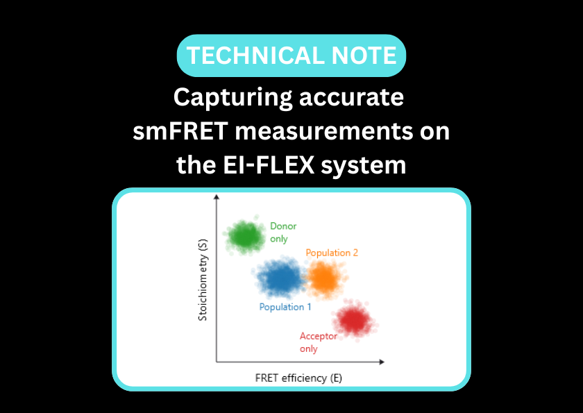
Single-molecule Förster resonance energy transfer (smFRET) and fluorescence correlation spectroscopy (FCS) offer highly complementary insights by moving beyond ensemble measurements to observe individual molecular events. These high-resolution approaches operate with single-molecule sensitivity, capturing the true heterogeneity of samples and identifying rare species present at low percentage abundance.
Single-molecule analysis of protein aggregation: Overcoming the challenges of ensemble measurements
- Detect early-stage oligomers, separating them from larger aggregates and classifying various stages of aggregation
- Characterise protein conformational changes that promote aggregation
- Capture protein heterogeneity in samples, with the ability to resolve multiple distinct populations simultaneously
- Track aggregation kinetics in real-time
- Identify self-association of therapeutics or interactions with formulation components or excipients
Understand the conformational changes that lead to aggregation, while detecting early dimers before they develop into large-scale fibrils
In the illustrated example below, single-colour FCS can be used to differentiate monomers from small aggregates and larger structures. Additionally, smFRET is ideal for understanding how proteins, such as therapeutic antibodies, can unfold and how this affects their propensity to aggregate.
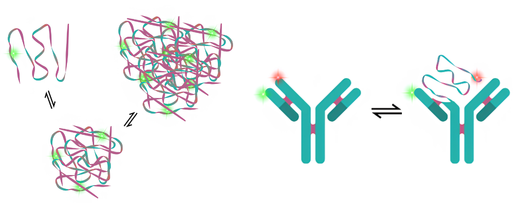
Figure 1 – smFRET reveals heterogeneity in pH-responsive DNA nanoswitches
a) At pH 7.6, the DNA nanoswitch is closed (high-FRET population)
b) At pH 7.8, there is a heterogeneous population of open and closed nanoswitches (low and high-FRET populations)
c) At pH 8.1, all DNA nanoswitches are open (low-FRET p`opulation)
Figure taken from: D’Rozario, F. et al. Electronic Actuation of Surface-Immobilized, pH-Responsive DNA Nanoswitches. ACS Appl. Mater. Interfaces 18, 18039–18048 (2026)
Protein aggregation papers and pre-prints featuring EI-FLEX data

Protein aggregation FAQs
How do single-molecule techniques differ from traditional Dynamic Light Scattering (DLS)?
While DLS provides an average size of all particles in a sample, it is easily biased by a few large dust particles or large aggregates, often masking smaller, toxic oligomers. Our single-molecule approach detects individual particles, allowing us to resolve distinct populations (monomers, dimers, and small oligomers) that DLS would average out.
Can I detect aggregation in messy samples like serum or cell lysates?
Yes. Because these techniques use fluorescence, biomolecules of interest can be separated from the rest of the solution, minimising interference from the surrounding environment.
Can this technology help predict long-term developability and shelf-life?
Absolutely. These techniques can detect reversible self-association (RSA) and early-stage dimerisation. Identifying these weak interactions early is a strong predictor of which candidates will eventually fail due to irreversible aggregation during long-term storage.
How can this technology visualise how excipients or stabilisers interact with my protein?
Fluorescence Cross-Correlation Spectroscopy (FCCS) can simultaneously track both a therapeutic protein and the excipient molecules (such as surfactants or polymers) in the same solution. By measuring the co-diffusion of these two colours, the percentage of the stabiliser that is physically bound to the protein surface can be quantified.
Can we distinguish between colloidal and conformational aggregation?
Yes. FCS tracks the physical size (colloidal clumping), while smFRET monitors the internal folding state (conformational misfolding). Combining these methods can determine if a protein is transiently sticking together or whether it has unfolded and exposed aggregation hot spots.
Is it possible to identify different polymorphs of misfolded proteins?
smFRET and FCS can distinguish different aggregate ‘strains’ (e.g., different polymorphs of Alpha-synuclein or Beta-amyloid) as they have distinct FRET efficiency signatures, hydrodynamic radii and propensities to aggregate, allowing researchers to fingerprint and categorise them, even if they are present within the same sample.
Can we monitor the effect of drug candidates on disaggregating existing fibrils?
Yes. By labelling aggregates and adding a potential drug, FCS can monitor disaggregation via changes in brightness and diffusion speed of the aggregate. This provides a real-time, quantitative readout of whether a drug is successfully breaking down aggregates or preventing new ones from forming.
How does the technology help with high-concentration viscosity challenges?
At high concentrations, antibodies can form transient networks that lead to high viscosity. FCS can measure the local viscosity and rotational dynamics of these molecules, helping to clarify whether a viscosity problem is caused by specific, targeted interactions or general crowding. The impact of changing pH, the salt concentration, or the excipient profile can also be measured.
"It has opened up new avenues of research because of the combined FCS, FCCS and single-molecule FRET capabilities. The high performance, stability and precision of the EI-FLEX instrument have really been a game-changer for experiments where sensitivity to picomolar concentration of biomolecules really matters."
Dr. Steven Quinn, University of York

Other resources you might be interested in