To perform a single-molecule Förster Resonance Energy Transfer (smFRET) experiment, biomolecules of interest must be labelled with a dye pair that can undergo FRET. The choice of dyes and their placement has a profound impact on the resultant data and therefore must be considered carefully when designing smFRET experiments.
The technical note will guide you through these practical considerations and provide recommended protocols to help you prepare for your own smFRET experiments.
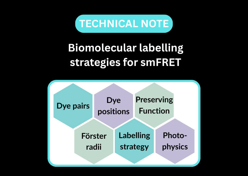
Overview of this technical note:
- Discussion of various commercially available dye pairs and ideal characteristics for FRET
- Methods for site-specific labelling for proteins and nucleic acids
- Important considerations that influence how a dye pair may behave in different biomolecular environments or distances from one another

Figure 1 – Excitation and emission spectra for Cy3 and Cy5 fluorophores
Excitation spectra are indicated by dashed lines; emission spectra are indicated by solid lines. Hashed areas show overlap between Cy3 emission and Cy5 excitation wavelengths, where FRET can occur.
Note – spectra are illustrative, not exact.